Introduction
The study of cranial morphometry holds pivotal significance in anthropology and basic medical research, aiding in species identification and determination of sex from skeletal remains. Cranial osteometry further facilitates the understanding of neurological and skeletal pathologies' origins and assists in devising surgical procedures and approaches.1 The skull, comprising bones interconnected at sutures, forms the skeletal framework of the head. Within the base of the skull lie various openings termed foramina, with the foramen magnum representing the largest aperture situated in the occipital bone's anteromedian position.2
The foramen magnum serves as a conduit for several vital structures, including the medulla oblongata with its meninges, the vertebral artery, anterior and posterior spinal arteries, and the accessory nerve.3 Its anterior margin is delineated by the basilar part of the occipital bone, while the condylar part of the occipital bone forms the lateral margins. The posterior margin comprises the squamous part of the occipital bone.1 On each side of the foramen magnum, there are two convex kidney-shaped condylar facets that articulate with the first cervical vertebra, forming the atlantooccipital joint. Internally and superiorly to the condylar processes lie the hypoglossal canals, connecting the internal posterior cranial fossa to the external cranial base, providing passage for cranial nerve XII.4
Assessment of the parameters of the foramen magnum and its index holds significant utility in craniovertebral and cervical spine surgeries. These findings offer invaluable guidance to neurosurgeons, particularly in executing lateral transcondylar surgical approaches to access lesions in the middle and posterior parts of the cranial base. The measurement of the largest anteroposterior diameter aids in achieving greater contralateral surgical exposure during transcondylar approaches, particularly in condylar resection. Additionally, a larger width of the foramen magnum correlates with a higher grade of cerebellar tonsillar herniation. Understanding the morphological shape of the foramen magnum is crucial for surgeons, as an ovoid shape can present challenges in traversing its anterior portion. Anatomical variations in the foramen magnum can significantly impact surgeries such as the repair of posterior inferior cerebellar artery aneurysms, decompression of the foramen magnum, and resection of meningiomas located within the foramen magnum.5 Furthermore, evaluations of the foramen magnum are pivotal not only for determining the most appropriate surgical techniques but also for obtaining valuable data in forensic medicine, aiding in the estimation of unknown sex and facilitating identity determination.6
The aim of present study is to create baseline data of anthropometric measurements on foramen magnum and cranium and to correlate foramen magnum index and cranial index. Through a comprehensive examination of the anatomical features of foramen magnum and cranium, we seek to unravel the complexities of skull morphometry and its clinical significance in the realm of forensic, radiology, orthopedics and neurosurgery.
Materials and Methods
The current study was conducted at the Department of Anatomy, Shri Guru Ram Rai Institute of Medical and Health Sciences, Dehradun. A total of 50 adult dry skulls, of unknown sex and age, were examined. The study was conducted from October 2023 to March 2024. Exclusion criteria encompassed damaged skulls and those displaying abnormalities. The study was conducted by using an vernier caliper and a spreading caliper.
Approval from the institutional ethics committee was obtained before commencing the study.
Parameters measured:
Anteroposterior (Sagittal Diameter of foramen magnum (FM in mm
Transverse Diameter of the foramen magnum (FM in mm
Foramen Magnum Index
Maximum Cranial Length in mm
Maximum Cranial Breadth in mm
Cranial Index
Types of Cranium
Following were the various landmarks used for the measurements of parameters:
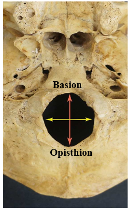
Basion: Median point on the anterior margin of the foramen magnum (Figure 1).
Opisthion: Median point on the posterior margin of foramen magnum (Figure 1).
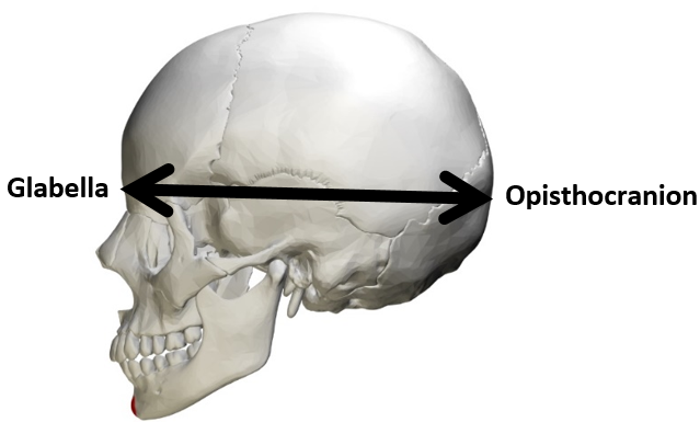
Glabella: Most prominent point on the middle of frontal bone between the two superciliary arches (Figure 2).
Opisthocranion: Most posterior point on the skull above the external occipital protuberance (Figure 2).
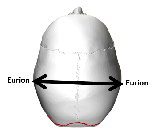
Eurion: Most lateral point on the either side of skull (Figure 3).
Measurements taken were:
Anteroposterior / Sagittal Diameter(APD of FM: From Basion to Opisthion (Figure 1)
Transverse Diameter (TD of FM: Maximum diameter in transverse plane (Figure 1)
Foramen magnum index (FMI = (TD/APD ×100
Maximum Cranial Length (CL): This is the distance between the glabella and opisthocranion (Figure 2).
Maximum Cranial Breadth (CB): it is the linear distance measured between the eurion points located on both parietal bones (Figure 3)
Cranial Index (CI = CB / CL × 100
The data collected from the dimensions of foramen magnum and cranium were statistically analyzed. Statistical analysis was done with statistical package for social sciences (SPSS Inc; Chicago, IL, version 20.0 for Windows). T-test was used for the analysis and significance of the p-value in this study.
Results
Table 1
Mean and standard deviation of various parameters
The average anteroposterior (AP) diameter of the foramen magnum was determined to be 33.33 mm, while the transverse diameter (TD) measured at 28.93 mm. Calculating the foramen magnum index (FM index) by dividing TD by AP diameter yielded a value of 83.85 mm. Additionally, the mean length of the cranium was recorded as 180.87 mm, with a mean breadth of 130.71 mm. The cranial index, obtained by dividing the breadth by the length of the cranium, was found to be 71.34 mm (Table 1).
Table 2
Correlation between parameters offoramen magnum and cranium
|
Variables |
Pearson's r |
p-value |
|
|
Foramen magnum Sagittal diameter |
Cranial length |
0.891 |
.001 |
|
Foramen magnum Transverse diameter |
Cranial breadth |
0.908 |
.001 |
|
Foramen magnum index |
Cranial index |
0.200 |
0.163 |
On comparison, a strong positive correlation (r= 0.891) was observed between the sagittal diameter of the foramen magnum and cranial length, demonstrating statistical significance (p-value < 0.05). Similarly, a strong positive correlation (r= 0.908) was identified between the transverse diameter of the foramen magnum and cranial breadth, also exhibiting statistical significance (p-value < 0.05). Conversely, when comparing the indices of the foramen magnum and the cranium, a weak correlation (r = 0.200) was detected, which was not statistically significant (p-value > 0.05) (Table 2).
Table 3
Comparison of anteroposterior diameter of FM, Transverse diameter of FM and FM Index of present study with previous studies
|
Author |
Anteroposterior Diameter of FM (mm) |
Transverse Diameter of FM (mm) |
Foramen Magnum Index |
|
Sharma et al1 (2019) |
34.44 |
30.46 |
88.44 |
|
Bharti et al5 (2021) |
Males: 30 |
Males: 26.1 |
Males: 87.33 |
|
Females: 29 |
Females: 25.03 |
Females: 85.54 |
|
|
Chandekar et al3 (2017) |
Males: 36.23 |
Males: 29.06 |
Males: 80.19 |
|
Females: 31.5 |
Females: 27.41 |
Females: 87.01 |
|
|
Singh et al7 (2019) |
33.57 |
27.49 |
82.09 |
|
Present study |
33.33 |
28.93 |
83.85 |
Table 4
Comparison of maximum cranial length, maximum cranial breadth and cranial Index of present study with previous studies
|
Author |
Maximum cranial length (mm) |
Cranial breadth (mm) |
Cranial Index |
|
Howale et al8 (2012) |
171.1 |
129.8 |
75.49 |
|
Senol et al9 (2019) |
172.20 |
139.15 |
81.59 |
|
Singh et al7 (2018) |
Males: 186.30 |
Males: 144.58 |
Males: 77.71 |
|
Females: 173.61 |
Females: 137.63 |
Females: 79.35 |
|
|
Present study |
180.87 |
130.71 |
71.34 |
Discussion
The foramen magnum serves as a pivotal junction between the skull and the vertebral column, holding considerable anatomical significance due to its proximity to vital structures including the brain, spinal cord, accessory spinal nerves, and vertebral arteries. Its morphological variability within the skull reflects evolutionary adaptations. A thorough understanding of the anatomy of the foramen magnum is essential for comprehending the pathophysiology of various craniovertebral junction disorders and for guiding surgical interventions.2 Cranial anthropometry holds increasing significance for anatomists, anthropologists, and plastic surgeons, facilitating detailed analysis and understanding of cranial structures and their variations.8
The current study presents a strong positive correlation between the length of cranium and the sagittal diameter of foramen magnum. Similarly, correlation between the breadth of cranium and the transverse diameter of foramen magnum also showed a strong positive correlation This shows that the size of foramen magnum increases with an increase in the size cranium.
In the present study, the mean foramen magnum index was determined to be 83.85. This contrasts with Sharma et al's findings, which reported an average index of 88.44. Bharti et al observed a mean index of 87.33 in male skulls and 85.54 in female skulls. Chandekar et al on the other hand, found differing averages with 80.19 in male skulls and 87.01 in female skulls. Singh et al estimated the mean foramen index to be 82.09. These variations highlight the diversity in foramen magnum morphology across different studies and populations (Table 3). In the present study,the mean cranial index was recorded as 71.34. This contrasts with Howale et al's findings, who calculated a mean index of 75.49. Senol et al reported a higher average index of 81.59. Singh et al found differing averages between genders, with 77.71 in males and 79.35 in females. Nair et al estimated the mean cranial index to be 76.67. These variations underscore the diversity in cranial morphology across different studies and populations (Table 4).
The morphometric analysis of the foramen magnum and cranium somewhat aligns with previous literature, indicating similarities in their measurements and supporting existing findings.
Limitation
The findings of the current study lead to the conclusion that variations in the shape of the foramen magnum have clinical as well as radiological significance. However, further exploration of this method is warranted, particularly through increasing the sample size and utilizing alternative methods for measurements such as radiology-based study. These approaches could enhance the accuracy and reliability of cranial morphometry in forensic and various other medical disciplines.
Conclusion
All parameters, except for indices, were found to be statistically significant, with a strong positive correlation observed among them. This suggests that as the size of the cranium increases, so does the size of the foramen magnum. These findings hold potential implications for various medical disciplines, including neurosurgery, orthopaedic surgery, radiology, forensic science, anatomy, and anthropology. It will provide important information to clinicians and neurosurgeons to approach the cranial base with maximum safety and minimal mortality and morbidity.