- Visibility 26 Views
- Downloads 4 Downloads
- DOI 10.18231/j.ijcap.2024.032
-
CrossMark

Study of nutrient foramina in dry adult femur bones in a medical college from north India
- Author Details:
-
 Yasmeena Akhter
-
Kaneez Fatima
-
Ghulam Mohammad Bhat
-
Bashir Ahmad Shah
-
Zameer Ali
Introduction
The femur is the longest bone of the body and has three main parts upper end, shaft and lower end. The upper end consists of the head, neck, greater and lesser trochanters, intertrochanteric line and intertrochanteric crest. The shaft of femur is cylindrical in shape, convex in front and narrowest in the middle. The femoral shaft is divided into upper 3rd having four borders and four surfaces, middle 3rd having three borders and three surfaces and lower 3rd having four borders and four surfaces. Linea aspera is ridge on middle of posterior border having inner lip, outer lip and an intermediate area.[1]
Nutrient foramina form important landmarks on human bones as they form portal of entry for nutrient artery. Nutrient artery is an important source of blood supply for a growing bone2. Long bones are supplied by a nutrient artery that enters individual bones obliquely through a nutrient foramen. This foramen in most cases is located away from the growing end hence the derivation of the axiom that foramen ‘seek the elbow and flee from the knee’. This is because one end of the limb bone grows faster than the other.[2], [3] Henderson (1978) reported that their position in mammalian bones are variable and may alter during growth.[4] The topographical knowledge of the nutrient foramen is useful in certain operative procedures to preserve the circulation.[5], [6] Therefore it is important that the arterial supply is preserved in free vascularized bone grafts so that the osteocytes and osteoblasts survive.[7] It is well known that one of the causes of delayed union or nonunion of fracture is lack of arterial supply.[8] The morphological knowledge of nutrient foramina is significantly important for orthopedic surgeons undertaking an open reduction of a fracture to avoid injuring the nutrient artery and thus lessening the chances of delayed or non-union of fracture.[8] The external opening of the nutrient canal, usually referred to as the nutrient foramen, has a particular position for each bone.[9] Two well-known factors may affect nutrient foramen position. These are growth rates at two ends of the shaft and bone remodeling.[4] Addressing the growth rates and bone remodeling, the bones of different age groups need to be studied. It has been suggested that the pull of muscle attachments on the periosteum explained certain anomalous nutrient foramina directions.[10] Nutrient arteries which are the main blood supply to long bones are particularly vital during the active growth period and at the early phases of ossification.[11] The nutrient artery of femur may arise from the medial circumflex femoral artery or from any artery parallel to the diaphysis. The earliest worker who described the direction of nutrient canal and their angle of entry was Havers (1691).[12] It was Berard (1835) who was first person to correlate ossification with bone growth and ossification with direction of canal.[13] Humphery (1861) also worked on direction and obliquity of nutrient canal and stated that direction of the canal is away from growing end.[14] The nutrient foramen is initially horizontal in direction and vessels divide into ascending and descending branches are almost at right angles to each other. In a few cases the role of vascular necrosis is pointed out.[15] In clinical practice the knowledge of growing ends is important. In young age injury or infection of the growing end may result in stunting of bone.[16] In orthopedics the development of new transplantation and resection techniques requires detailed data on the blood supply to the long bones and association with the areas of bone supplied.[11]
Detailed information of nutrient foramen has a great importance in bone transplant and resection techniques and other orthopedic surgical procedures involving femurs. As there is racial, genetic and ethnic variation amongst the human femur bones, the present study was conducted to find out the exact location, number, size, and direction of nutrient foramina in human adult femurs.
Materials and Methods
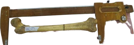
The study was conducted in the department of Anatomy of a medical college in central district of Kashmir after ethical clearance from institutional ethical committee. The material for present study comprised of 100 dry femur bones taken from department of Anatomy and included bone of either sex or side. Bones with gross deformities like evidence of any previous trauma, skeletal disorders, osteoarthritic changes or bones with ill-defined nutrient foramina and those having foramina at ends were excluded. After determining the side of femur bones, the length of the bones was measured by the osteometric board. The presence of foramina was identified by the presence of a well-marked groove leading it to a canal, which has slightly raised edges at the commencement of the canal. The nutrient foramina were studied with regards to number, direction from growing end and distribution in relation with length of femur. Keen observation was made for the direction of nutrient foramen by using a magnifying hand lens and then a thin stiff wire was passed through the foramen to confirm its direction. Nutrient foramen in each bone was encircled using a black marker pen. Size of the nutrient foramen was determined by using hypodermic needles of 20G and 22G. Nutrient foramen accepting the needle 20G were considered as larger size, while as nutrient foramen accepting the needle 22G were considered as smaller size. All the dry adult human lower limb long bones were digitally photographed, and variations about direction, location and number of nutrient foramina were noted and photographed using digital camera. The position of all the nutrient foramina was determined by calculating a Foraminal Index (Fl) by applying the Hughes formula FI = DNF / TL× 100 whereas DNF is the distance from the proximal end of the bone to the nutrient foramen measured by Vernier caliper and TL is total length of femur using osteometric board. The observations were recorded, and the data was analyzed statistically.
Results
Out of 100 femurs, 56 femurs were right sided and 44 were left sided. Single foramina were noted in 46 right sided and 29 left sided femurs while as double foramina were found in 10 right sided and 15 left sided femurs. Overall, single foramina were seen in 75% of femurs. The distribution as per the position of nutrient foramen in Femurs in shown in [Table 1].
|
Single |
Double |
||||
|
No. |
%age |
No. |
%age |
||
|
LA |
ML |
68 |
90.7 |
19 |
76.0 |
|
LL |
6 |
8.0 |
6 |
24.0 |
|
|
MS |
1 |
1.3 |
0 |
0.0 |
|
|
LS |
0 |
0.0 |
0 |
0.0 |
|
|
PS |
0 |
0.0 |
0 |
0.0 |
|
|
AS |
0 |
0.0 |
0 |
0.0 |
|
|
Total |
75 |
100 |
25 |
100 |
It was noted that in all the right (56) sided femurs, the direction of the nutrient foramen was away from the growing ends while as among left sided femurs only one femur had the direction of foramen towards growing end. Overall, among 100 femurs 99% had direction of the nutrient foramen opposite to the growing end as shown in [Figure 1].


Among the right sided femurs 52 foramens were patent and 4 foramens were non-patent while as in the left sided femurs, the foramen was patent in 43 femurs and only 1 left sided femur with two non-patent foramens was found. Overall, 5% nutrient foramens were noted to be non-patent and 95% of the foramen were found to be patent.
|
Parameter |
Mean |
SD |
Range |
95% CI |
|
Total Length |
38.9 |
2.51 |
34-44 |
38.3-39.5 |
|
Length from the proximal end |
16.1 |
5.21 |
7-26 |
15.1-16.9 |
|
Foramina Index |
40.8 |
12.62 |
17.9-60.5 |
38.2-43.3 |
All right sided femurs and 32 left sided femurs had foramina which were found to be large size while as 12 left sided femurs had small sized foramina. Overall 88% femurs had large sized foramina.


Discussion
It is well known that one of the causes of delayed union or non-union of fracture is lack of arterial supply.[17] The biologic process of repair of a traumatic or surgically induced fracture has been described as developing slowly or not at all.[16] The morphological knowledge of nutrient foramina is significantly important for orthopedic surgeons undertaking an open reduction of a fracture to avoid injuring the nutrient artery and thus lessening the chances of delayed or non-union of the fracture.[7] The external opening of the nutrient canal, usually referred to as the nutrient foramen, has a particular position for each bone.[18] It is generally agreed that the vessels which occupy the nutrient foramen are derived from those that took part in the initial invasion of the ossifying cartilage, so that the nutrient foramen was at the site of the original center ofossification.[18] In our study mean femur length was 38.9cm with SD of 2.5cm which is comparable with Indian population from different states as reported by Parmar A et al.[15] Parmar A reported mean length of femur in Maharashtra, Tamil Nadu, North India, South India and Rajasthan populations as 40.8 cm, 42.2 cm,43.6cm 41.8 cm and 40 cm respectively. In the present study, single nutrient foramen was found in 75% of the femur like in the studies by Longia GS et al[12] (85% of 200 long bones of limbs), Pereira GA Metal et al[13] (88.5% of 174 bones), Solanke KS et al[14] (92 of 100 bones) and Roul B and Goyal M et al[19] (94.6% of 37 bones). In the present study, two nutrient foramina were found in 25% of femurs. Seemaet al[20] observed single nutrient foramen in femur in 48.85% of cases and double nutrient foramina were found in 47.71% of cases. Mysorekar VR[5] (1967) had reported that out of 180 femora, ninety-three (more than 51%) had more than one nutrient foramina. The double nutrient foramen of the femur was observed in 30% by Kalyanasundaram et al, [21] 33% by Vinay and Mangala Gowri[22] and 47.7% by Murli Manju et al.[23] In a study by Murli Manju et al.[23] 47.7% of the femora had a single nutrient foramen. The double foramen was observed in 44.2% of the cases, triple foramen in 3.5% and the foramen were found to be absent in 4.6% of the femora. In the present study, all the nutrient foramina are directed upwards, and the majority were of large size similar to study done Singh A K[16] et al. In our study 85% of femurs had nutrient foramina on Linea aspera similar to that reported by Gumusburun E et al[24] (65% of the 188 foramina were present on the Linea aspera and its lips).Prashanth KU et al[23] reported that Sixty-six femora showed the foramina on the Linea aspera (LA), 37 at the medial lip of Linea aspera (ML), 5 had foramen at the lateral lip of Linea aspera (LL), 16 at medial surface (MS) and 1 each at lateral (LS) and popliteal surfaces (PS). Collipal E et al[25] investigated the 140 dry femora for the location and number of diaphysis foramina. The nutrient foramen of the Femur was on the Linea aspera in 72.5%, on medial surface in the 21.25% and on the lateral surface of the diaphysis of the bone in 6.25% like our study.
Conclusion
In the present study, morphology and topography of nutrient foramen in femurs were studied and the observations made were tabulated and statistically analyzed. The findings of the present study were compared with that of the results of the previous studies and with the standard textbooks of anatomy and they were found markedly similar. The morphometric study of the nutrient foramen with regards to the number, location and position is of great importance for clinicians, radiologists and vascular surgeons as exact knowledge of position and distribution of nutrient foramina in bones is important to avoid damage to the nutrient vessels during surgical procedures like vascularized free grafts, joint replacement therapy, fracture repair, bone grafts, and in medico-legal cases and so surgeons should be mindful of soft tissue in the foraminal area during surgical procedures.
Source of Funding
None.
Conflict of Interest
None.
References
- K Garg, PS Mittal, M Chandrupatla. . B D Chaurasia’s Human Anatomy 2019. [Google Scholar]
- VR Mysorekar, A Nandedkar. Diaphyseal nutrient foramina in human phalanges. J Anat 1979. [Google Scholar]
- SM Patake, VR Mysorekar. Diaphyseal nutrient foramina in human metacarpals and metatarsals. J Anat 1977. [Google Scholar]
- RG Henderson. The position of the nutrient foramen in the growing tibia and femur of the rat. J Anat 1978. [Google Scholar]
- VR Mysorekar. Diaphyseal nutrient foramina in human long bones. J Anat 1967. [Google Scholar]
- DP Green. . Operative hand surgery 1988. [Google Scholar]
- H Joshi, B Doshi, O Malukar. A study of the nutrient foramina of the humeral diaphysis. Natl J Integrated Res Med 2011. [Google Scholar]
- C Havers. . Osteological Nova: some new observation of the bones and the parts belonging to them 1691. [Google Scholar]
- GM Humphery. Observations on the growth of long bones and stumps. Medico Chir Trans 1861. [Google Scholar]
- AK Datta. . Essentials of Human Osteology 2005. [Google Scholar]
- JG Craig, D Widman, MV Holsbeeck. Longitudinal stress fracture: patterns of edema and the importance of the nutrient foramen. Skeletal Radiol 2003. [Google Scholar]
- GS Longia, ML Ajmani, SK Saxena, RJ Thomas. Study of diaphyseal nutrient foramina in human long bones. Acta Anat (Basel) 1980. [Google Scholar]
- GAM Pereira, PTC Lopes, AMPV Santos, FHS Silveria. Nutrient foramina in the upper and lower limb long bones of Southern Brazilian adults. Int J Morphol 2011. [Google Scholar]
- KS Solanke, R Bhatnagar, R Pokhrel. Number and position of nutrient foramina in humerus, radius and ulna of human dry bones of Indian origin with clinical correlation. OA Anat 2014. [Google Scholar]
- A Parmar, P Maheria, K Shah. Study of nutrient foramina in human typical long bones of lower limb. Nat J Clin Anat 2019. [Google Scholar]
- AK Singh, R Kumari. Evaluation of nutrient foramina of the dry adult femur bone of north Indian population. Acad Anat Int 2019. [Google Scholar]
- P Lacroix. . The organization of bones 1951. [Google Scholar]
- CG Payton. The position of the nutrient foramen and direction of the nutrient canal in the long bones of the madder-fed pig. J Anat 1934. [Google Scholar]
- B Roul, M Goyal. A study of nutrient foramen in long bones of inferior extremity in human being. Int J Adv Res 2015. [Google Scholar]
- Seema, P Verma, A Mahajan, D Gandhi. Variation in the number and position of nutrient foramina of long bones of lower limb in North Indians. Int J Anat Res 2015. [Google Scholar]
- M Kalyanasundaram, D Backiaraj, R Shalini, R Manoranjitham. Morphometric study of nutrient foramen in the long bones of lower limb. Int J Anat Res 2017. [Google Scholar]
- G Vinay, SRM Gowri. Anatomical study of the nutrient foramen of lower limb long bones in South Indian population. Indian J Clin Anat Physiol 2017. [Google Scholar]
- B Murlimanju, K Prashanth, LV Prabhu, GK Chettiar, MM Pai, K Dhananjaya. Morphological and topographical anatomy of nutrient foramina in the lower limb long bones and its clinical importance. Australas Med J 2011. [Google Scholar]
- E Gumusburun, F Yucel, Y Ozkan, Z Akgun. A study of the nutrient foramina of lower limb long bones. Surg Radiol Anat 1994. [Google Scholar]
- E Collipal, R Vargas, X Parra, H Silva, SM Del. Diaphyseal Nutrient Foramina in the Femur, Tibia and Fibula Bones. Int J. Morphol 2007. [Google Scholar]