Introduction
Sacrum is a large triangular bone formed by the fusion of five sacral vertebrae. In an articulated pelvis it forms the posterior boundary of the pelvic cavity. It is placed obliquely like a wedge in between two hip bones. It forms an angle with rest of the vertebral column known as the sacro-vertebral angle, which is varies between 128-160 degree with an average of 140 degree in male and 137 degree in the female. The sacrum consist of a base, an apex and the four surfaces, pelvic, dorsal, and a pair of lateral. The sacrum is divided by the rows of foramina into a median portion traversed by the fusion of the transverse by sacral canal and a pair of lateral masses formed by the fusion of the transverse processes posteriorly with the costal elements anteriorly. The base is directed upwards and forwards and consist of an articular part and a non-articular part, the articular part is formed by the upper surface of the body of first sacral vertebra. The anterior margin of the articular part of the base projecting forwards forming a prominent margin known as the sacral promontory. The non-articular part of the base projects lateral wards on either side of the articular part, known as ala of sacrum. The ala of sacrum forms the upper surface of the lateral mass and is formed by the fusion of transverse process and costal elements. The vertebral foramen is large and triangular. It lies behind the body of S1 vertebra and leads into the sacral canal. The pedicles are short and directed backwards and laterally. The laminae are directed downwards, backwards and medially. Where the meet, the spinous process is represented by spinous tubercle. The pelvic surface is concave from above downwards and also from side to side. Due to the oblique position of the sacrum its pelvic surface is directed downward and forwards. There are four pairs of the pelvic sacral foramina communicate through intervertebral foramina with sacral canal. There are four transverse ridge which indicates the line of fusion between sacral vertebrae. The bar of bone intervening between two sacral foramina of the same side represents the costal elements. Lateral to the anterior sacral foramina on each side, costal elements unite with one another to form the lateral mass. The dorsal surface of sacrum is rough, irregular and convex and is directed backward and upwards. In the median plane it is marked by median sacral crest with 3-4 spinous tubercles representing the fused spines of the upper four sacral vertebrae. Below the 4th spinous tubercle there is an inverted U shaped gape called sacral hiatus. The articular surface formed by the costal elements, articulate with the articular surface of the hip bone. Apex of the sacrum is formed by the inferior surface of the body of S5 vertebra. It bears an oval facet for coccyx. It has long been customary among anatomists, anthropologists and forensic experts to identify sex of skeletal material by metric and non-metric observations. Sacrum is an important bone for this purpose, both as an individual bone and a part of pelvis. It has an applied importance in determining the sex with the help of measurements carried out upon it. Metrical study of sacrum for the identification of sex have been carried out by various authors.1, 2, 3
Materials and Methods
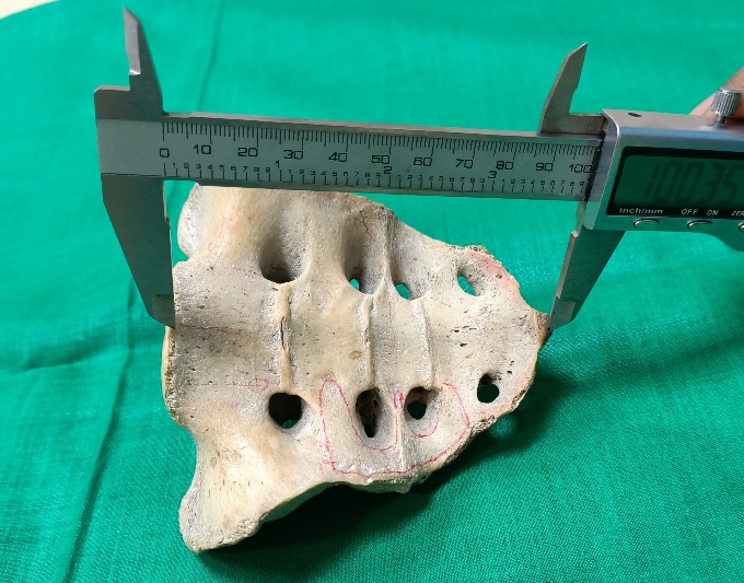
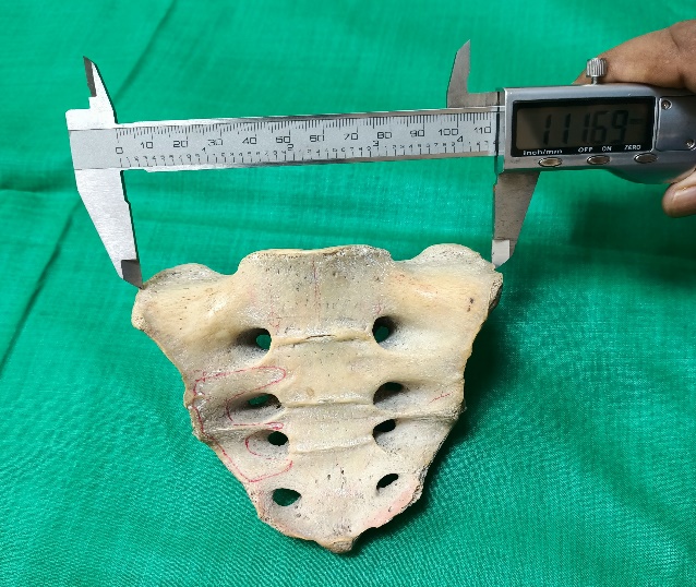
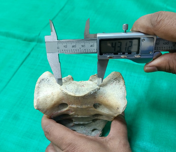
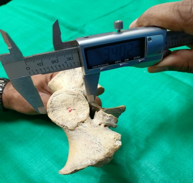
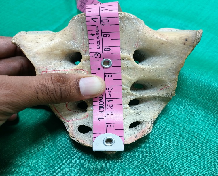
The present study consist of 110 adult sacra of known sex (62 male and 48 female). These were collected from department of Anatomy, Indira Gandhi Institute of Medical Sciences, Patna, Bihar and also from other medical college from August 2018 to September 2019. The sacra were measured with a sliding digital caliper, flexible steel measuring tape, scale and dividers.
The following measurement were taken:-
Maximum length of sacrum
Curved length of sacrum
Maximum breadth of sacrum
Antero-posterior diameter of the body of first sacral vertebra-
Transverse diameter of the body of first sacral vertebra
Maximum length of articular surface
By using above measurements the following indices were calculated:-
Results
Table 1
Sacral parameters
Table 2
Indices of sacrum
In the present study, 110 sacra are examined in view of the sacral dimorphism of sacrum. The mean straight length is 104.55 mm and 94.66 mm in male and female sacrum respectively. The mean curve length of sacrum is 112.03 mm and 103.98 mm in male and female respectively. The mean width in male sacra is 101.53 mm and in female sacra is 105.67 mm. The mean transverse diameter of body of 1st sacral vertebra is 46.53 mm and 40.85 mm in male and female sacra respectively. The mean antero-posterior diameter of body of 1st sacral vertebra are 29.89 mm and 27.73 mm in male and female sacra respectively. The mean length of auricular surface are 56.08 mm and 54.77 mm in male and female sacra respectively. The mean sacral index is 97.66 and 112.12 in male and female sacra respectively. The mean Curvature index is 93.28 in male and 91.21 in female. The mean corpobasal index is 45.85 in male and 43.46 in female. The mean Index of the body of 1st sacral vertebra is 64.38 in male and 67.68 in female.
Discussion
In the present study 110 adult human sacrum (62 male and 48 female) were studied in the department of Anatomy, Indira Gandhi Institute of Medical Sciences, Patna. Mean straight length of Sacrum were 104.55±7.53 mm and 94.66±7.89 mm in male and female respectively; mean width of sacrum were 101.53±5.80 mm and 105.67 mm in male and female respectively. These findings are in agreement with the yadav et.al.4 in which ventral straight length was 104.7±5.94 mm in male and 92.6±6.1 mm in female, maximum breadth was 102.93±4.83 mm in male and 104.77±6.48 mm in female.
The mean value of transverse diameter of body of 1st sacral vertebra was 46.53 mm in male and 40.85 mm in female and antero-posterior diameter was 29.89 mm and 27.73 mm in male and female respectively. These finding are in agreement with Mazumdar S et al (2012)5 who obtained mean transverse diameter of body of 1st sacral vertebra to be 41.6±8.5 mm in male and 39.7±52 mm in female while mean antero-posterior diameter was 29.48±3.8 mm and 27.9±2.7 mm in male and female respectively.
In this study the mean length of auricular surface was 56.08 mm and 54.73 mm in male and female respectively, this observation was very close to study done by Mishra et al (2003)6 in which mean length of auricular surface was 62.54 mm and 54.57 mm in male and female sacra respectively.
The mean sacral index was 97.66 in male and 112.12 in female. This is similar to the reported by earlier workers namely Patel MM et al (2005)7 which was 96.25±4.6 and 113.25±5.74 in male and female respectively.
In the present study, the mean curvature index in males and females were 93.28±2.37 and 91.21±4.22 respectively. Similar observation were reported by Raju and Singh8 who observed mean curvature index 92.77 in male and 88.5 in female.
In the present study, the mean corporobasal index were 45.85±3.39 in males and 43.46±3.12 in females. Similar observation were recorded by Maddikunta V et al9 who observed mean corporobasal index was 46.6±4.86 and 42.9±4.97 in male and female respectively.
In the present study, Index of the body of 1st sacral vertebra were 64.38±4.35 and 67.68±5.20 in male and female respectively. This observation is very similar to the study done by Shree Krishna et al10 in which Index of the body of 1st sacral vertebra was 64.33±6.43 69.40±6.9 in male and female respectively.

