Introduction
Binturong, often considered as the largest civet in the planet found in the forest remnant of South-east Asia. Common characteristic features of a Binturong is its shout legs, thick black body coat and prehensile tail, which makes it a dominant arboreal animal. Main threat to this omnivorous animal is its habitat loss due to degradation of forest or deforestation through logging and therefore has been appraised as vulnerable on the International Union for Conservation of Nature (IUCN) red list (Grassman et al., 2005, Kalita et al., 2020).1, 2 As it is standing on the verge of un-natural extinction, facts and statistics on the structural body features of binturong are few and far between. Even though gross morphological and morphometric researches on the different body regions of the Binturong have been done before; but studies on different features and morphometric analysis related to the forelimb of Binturong is not yet done. Since it’s the high time for human intervention to preserve this beauty of nature, every bits of information regarding the biology of this animal is very essential. This present study was conducted to get the baseline data about the two bones of forelimb in Binturong, which can play a predominant role in disease diagnosis, treatments and other applied fields of veterinary biology.
Materials and Methods
This current study was performed on the Shoulder blade, Arm bone of Binturong. Bones of the both left and right forelimbs were collected from the Aizawl Zoological Park, Mizoram with permission from the Department of Environment, Forest and Climate change, Government of Mizoram. Collected samples were macerated by boiling in hot water (Kalita et al., 2020, Simoens et al., 1994; Choudhary et al., 2015)3, 2, 4 and afterwards were treated with Hydrogen Peroxide (H2O2) solution then were sundried for several days (Bharti et al., 2021).5 This recent study was conducted to establish a baseline data on the morphological and morphometric features of the major forelimb bones of Binturong. Following parameters were measured:
Scapula
Total length: Maximum length of scapula was measured from the highest point in the dorsal border to the lowest point of the caudal border.
Spine length: Full length of the scapular spine.
Maximum total width of scapula.
Maximum length of supraspinous fossa.
Maximum length of infraspinous fossa.
Maximum width of supraspinous fossa.
Maximum width of infraspinous fossa.
Width of scapular neck.
Circumference of scapular neck.
Result & Discussion
Scapula
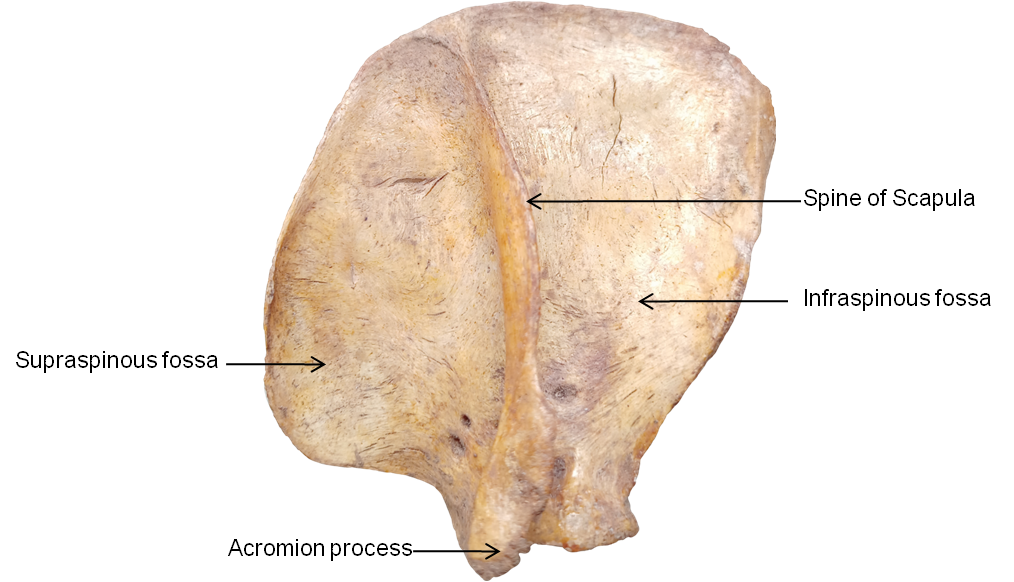
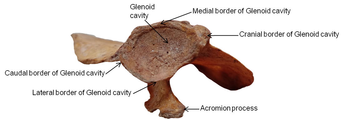
Shoulder blade or scapula of binturong was a flat almost square shaped bone with similar width in both dorsal and ventral end with a wide scapular neck, average circumference of which is around 8.04cm. However scapula was reported as a triangular flat bone in horse by Getty (1975)6 and by Choudhary et al., (2013)7 in Chital. Scapula of Binturong has 3 distinct border i.e. dorsal border, cranial broder and caudal border. Average maximum length and width of the scapula of Binturong was 8.81 cm and 6.56 cm, respectively (Table 1). Average maximum length and width of the scapula of Asiatic Lion was 18.28±0.90 cm and 10.83±0.36 cm, respectively. In the lateral surface of the scapula there was a prominent spine which divided the surface in to one large caudally present fossa i.e. infraspinous fossa and one comparatively smaller cranially present fossa i.e. supraspinous fossa (Figure 1). Caudal portion of the scapular spine crosses the scapular neck and thickened at the caudal most portions as acromion process, also reported in Black Bengal goat by Siddiqui et al., (2008)8 but in pig and horse the scapular spine subsides distally Liebich et al., (2004)9 (Figure 1). A small foramen was seen at the neck of the scapula perforating the ventral portion of the scapular spine. Tuber spine scapulae was not prominent as reported by Liebich et al., (2004)9 in carnivores (Figure 1). On the ventral side of scapula there was a shallow subscapularis fossa, analogous with finding of Liebich et al., (2004)9 in ruminats but contrary to the findings of Asiatic lion, Choudhary et al., (2013)7 in Chital and Siddiqui et al., (2008)8 in Black Bengal goat where they found it was markedly deep. Coracoid process is not properly defined as reported by Siddiqui et al. (2008)8 in Black Bengal, but it was well-developed in Elephants as described by Sarma et al., (2007). Caudal border carries the glenoid cavity, which is a deep cavity with a distinct notch on the ventral side of the scapula with an average depth of 1.19 cm (Table 1). There was no distinct glenoid notch reported on the Black Bengal Goat by Siddiqui et al., (2008)8 (Figure 1).
Humerus
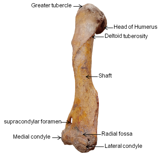
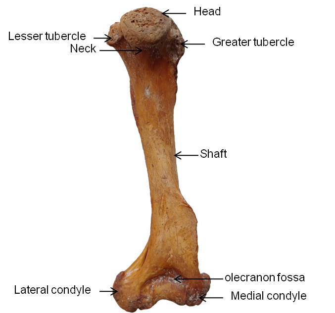
Humerus of Binturong is long, compact bone directed obliquely downward and backward which is comprises of a proximal and distal extremity joint by a less twisted cylindrical diaphysis or shaft (Figure 3). Similar findings were reported in Mithun by Talukdar et al., (2002)4 and in Blue Bull by Bharti et al., (2021),5 whereas the shaft was more twisted in Black Bengal Goat reported by Siddiqui et al., (2008).8 Average maximum total length of Binturong Humerus was 12.81 cm (Table 2). Basic segments of a Binturong humerus are proximal extremity, a shaft and a distal extremity. Proximal extremity of the humerus carries a caudally present oval shaped head: average maximum width of which is about 2.05cm (Table 2), a Neck: which separates the head from the shaft, a greater tubercle: present on the craniolateral aspect of the head, and a small lesser tubercle: present on the craniomedial aspect of the head (Figure 3). Maximum length of the proximal extremity was 2.52 cm (Table 2). These two greter and lesser tubercle are separated by a deep groove: Bicipital groove. Greater tubercle is comparatively large and form the lateral boundary of the bicipital groove, whereas the lesser tubercle is small and attached with the head, it forms the medial boundary of the bicipital groove. Similar findings were found in Mithun by Talukdar et al., (2002)4 and in Tiger by Tomar et al., (2014).10 Body or shaft of the humerus is the mid part of the bone which is twisted medio-laterally in its proximal two third and twisted cranio-caudally in its remaining parts. An oblique tricipital line arise from the caudal portion of the greater tubercle and ends in a small projection known as deltoid tuberocity: present on the cranial surface of the humerus, as in cat by Boyd et al., (2001) and in tiger by Tomar et al., (2014)10 in ox by Getty, R. (1975)6 in Mithun by Talukdar et al., (2002)4 (Figure 3). A small rough surface present on the end portion of the crest of lesser tubercle is homologues to the teres major in ruminants, also reported in carnivores by Liebich et al., (2004).9 Distal extremity of the Binturong humerus is consists of a supracondylar foramen, a radial fossa, a olecranon fossa, lateral and medial condyle and epicondyle. Prominent supracondylar foramen is present on the medial portion of the distal extremity of the humerus. A shallow radial fossa is present on the cranial aspect of the distal extremity, mainly on top of the lateral condyle (Figure 3). A comparatively deep olecranon fossa is present on the caudal aspect of the distal extremity (Figure 4). These findings are quite similar with the findings of Boyd et al. (2001) in cat and of Tomar et al., (2014)10 in tiger, but there was absence of supracondylar foramen in ox reported by Getty, R. (1975)6 and in Mithun reported by Talukdar et al., (2002).4 Average Maximum length and maximum width of the distal extremity of humerus of Binturong is 2.11 and 4.05 cm, respectively (Table 2).


