- Visibility 174 Views
- Downloads 14 Downloads
- DOI 10.18231/j.ijcap.2020.083
-
CrossMark
- Citation
Variations of the ulnar nerve and it’s clinical implications
- Author Details:
-
Prashant Munjamkar *
Introduction
As it emerges from the medial cord of the brachial plexus, the Ulnar nerve is also known as the musician's nerve, being the only branch with a root value of C8 and T1[1] and the most medial to the axillary artery. However, after the median nerve, it is the second most common nerve involved in compressive neuropathy. The posterior part of the medial epicondyle (sublime tubercle) of the humerus is grooved as it reaches the elbow joint.[2] It stretches between the two heads of the flexor carpi ulnaris into the forearm and passes along the flexor digitorum profundus under the flexor carpi ulnaris shield. The dorsal cutaneous nerve is delivered proximally to the wrist and supplies one and a half digits to the medial skin of the dorsum of the hand.
Over the flexor retinaculum, the ulnar nerve also gives off a small palmar cutaneous branch that supplies the skin over the hypothenar muscles. UN then descends on flexor digitorum profundus and runs to the hamate's hook laterally. It then gives off its superficial branch that delivers on the ulnar and palmar aspect, as well as palmaris brevis, one and a half digits. It then operates like the deep branch of the ulnar nerve between flexor digiti minimi and abductor digiti minimi. The deep branch follows the deep carpal arch and on the ulnar side of the wrist supplies both the interossei and the two lumbricals. The deep branch ends then, by getting adductor pollicis.[3], [4]
It should be noted that irregular UN structures and their uncommon interactions with neighboring nerves can be a complicating factor during surgical attempts to trigger nerve blockade.[5] UN variations are consistently found in the root or path of the distal branches. There is, however, a somewhat peculiar variation in the forearm ulnar nerve branching pattern that may be important to the diagnosis of unexpected and uncommon clinical conditions.[6] Although anatomical variations of the ulnar nerve have been documented in several studies, they most frequently relate to the anatomical branch or irregular contact of the ulnar nerve with other nerves in the arm or hand.[7], [8], [9] The present research was therefore conducted to determine the variance and possible clinical consequences of the ulnar nerve.
Materials and Methods
The present study was based on the dissection from the Department of Anatomy, Shri Shankaracharya Institute of Medical Sciences, Bhilai, Chhattisgarh, India of 40 upper limbs of formalin-embalmed human cadavers ranging in age from 55 to 70. Of the 40 upper extremities, 20 were left-sided and 20 were right-sided. After clearing the entire fascia, the axillary area of all the limbs was carefully revealed and the anatomy of the formation of the ulnar nerve from the medial cord of the brachial plexus was studied. In order to probe any ulnar nerve communications with neighbouring peripheral nerves, we continued with the dissection towards the anterior arm compartment. Meticulous observation, if any, of variant modes and/or irregular contact and all of its branches have been recognized.
Observations and Results
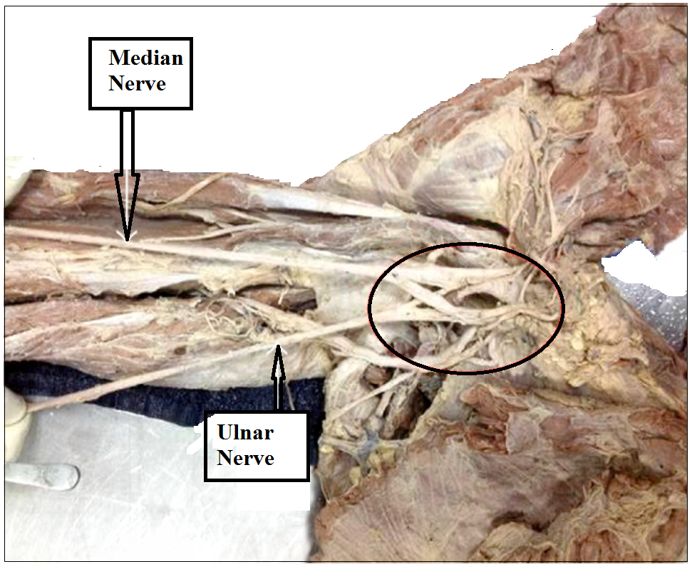
Four out of 40 upper limbs were presented with variations linked to the ulnar nerve and all four variations were found with an incidence of 10 percent in the right upper limbs. The irregular development of the ulnar nerve, with a significant input from the brachial plexus lateral cord ([Figure 1]), was observed unilaterally in two extremities. The contributing branch moved deep from the lateral to the medial side to the median nerve structure and entered the lateral part of the ulnar nerve. The formation of the ulnar nerve was similar to the median nerve formation. The nerve exhibited a regular course in the arm following its irregular creation. The ulnar nerve was also produced in 1 specimen by two roots that came from the medial cord and lateral cord, as shown in [Figure 2].


In one scenario, as shown in [Figure 3], the ulnar nerve had irregular contact with the neighbouring nerve, i.e. the medial cutaneous nerve of the forearm. In this case, in its medial aspect, the nerve gave off a transmitting branch of touch from the medial cutaneous nerve of the forearm. Before the latter perforated the medial intermuscular septum, the communicating branch sloped medially and joined the ulnar nerve pathway.

Discussion
Sensory innervation defects in the hand are uncommon.[10] Awareness of nerve variance is necessary and could clarify the patient's sudden sensory / motor loss or pain following surgery or trauma. Anatomical variations of the ulnar nerve consisting of irregular branches of the nerve or irregular communication between the ulnar nerve / its branches and other nerves of the hand have been documented in previous studies.[7], [8], [9] However, in the available literature, in terms of both its atypical formation and irregular contact with the neighbouring nerves, not much importance has been levied on the UN variations. In the present study, the peculiar aberrant formation of the UN was noted by the remarkable contribution of the lateral cord in three of the 40 upper limbs, which is correlated with the Emamhadi et al. study.[11] In addition, Guru et al[12] have suggested that variations in the formation of the ulnar nerve are found in 2 specimens where the lateral cord also contributes to the ulnar nerve. Goel et al[13] stated that the ulnar nerve emerges from the medial cord via two roots, which subsequently unite to form a single trunk. There are no such considerable variations in the present analysis. Iatrogenic injuries and trapping neuropathies are vulnerable to variations in the origin, course, and distribution of nerves.[14] It is therefore prudent to rule out its aberrant formation during diagnostic approaches to the seriousness of ulnar neuropathy.
Moreover, in the medial and lateral cords, irregular communication between the branches of the brachial plexus is frequently observed.[15] 60 per cent of cadavers had a communication in 200 dissected cadavers. Both proximal and distal communications in the dorsum of the hand have been identified, especially over the fourth digit.[16] Emamhadi et al[11] indicated that 6 brachial plexuses received ulnar nerve communication from the lateral cord. As found in our case, communication between the ulnar nerve and the medial cutaneous nerve of the forearm can only rarely be found. In this case, the nerve gave off a communicating branch in its medial aspect of communication from the medial cutaneous nerve of the forearm. Before the latter perforated the medial intermuscular septum, the communicating branch sloped medially and joined the ulnar nerve pathway. Similarly, Guru et al[12] stated that the ulnar nerve interacts with the radial nerve in one specimen and the medial cutaneous nerve of the forearm in another. A case in which the ulnar nerve was followed by the medial cutaneous nerve of the forearm within a typical fascial sheath was recorded by Das and Paul.[17] In nerve grafting and neurophysiological assessment for diagnosing peripheral nerve lesions, knowledge of certain variations affecting cutaneous nerves is beneficial.
The study outcomes mentioned clearly indicate that differences in the ulnar nerve are not unusual. The ulnar nerve is the second most common neuropathy to be involved in entrapment. During shoulder & elbow surgical procedures, these variations gain significance when the presence of an unexpected variation may lead to iatrogenic injury. The variations in the diagnosis of neurological syndromes can also present difficulties. In trauma to the upper limb, good knowledge of the distribution and variations of the ulnar nerve and its potential communication with the median nerve are valuable. Electrodiagnostic tests will detect all these variants. Electromyography is a valuable method in the diagnosis of abnormal arm, forearm and hand variations. In patients with ulnar nerve symptoms in which the clinician finds it difficult to determine the exact cause, there is a need to increase the understanding of the likelihood of anatomical variation and the frequency in which they occur and this can provide clues for a specific diagnosis of the possible cause of ulnar nerve trapping.
Conclusion
The typical sensory-motor activity may be affected by an irregular relationship of the ulnar nerve with its neighbouring nerves and its abnormal formation. Knowledge of the UN's anatomical variations is therefore important for clinicians to consider the nature of ulnar nerve neuropathy-related complications. The understanding of these variations along the usual pattern is often helpful during radiological procedures or operating on fractured patients or diagnostic therapy for interventional radiologists, orthopedics and neurologists to avoid untoward iatrogenic damage to the ulnar nerve.
Source of Funding
None.
Conflict of Interest
The authors declare no conflict of interest.
References
- PL Williams, M Dyson, S Standring, H Ellis, JC Healy, D Johnson. . Gray’s Anatomy 2005. [Google Scholar]
- A Arachchi, ZY Loo, H Maung, A Vasudevan. A rare anatomical variation between the radial and ulnar nerves in the arm. Int J Anat Variations 2013. [Google Scholar]
- G Ozguner, K Desdicioglu, S Albay. Connection between radial and ulnar nerves at high humeral level. Int J Anat Var (IJAV) 2010. [Google Scholar]
- CS Sinnatamby. . Last’s Anatomy: Regional and Applied 2011. [Google Scholar]
- S Stanescu, J Post, NA Ebraheim, AS Bailey, R Yeasting. Surgical anatomy of the radial nerve in the arm: practical considerations of the branching patterns to the triceps brachii. Orthop 1996. [Google Scholar] [Crossref]
- P Lama, BK Potu, KMR Bhat. High origin of dorsal branch of the ulnar nerve and variations in its branching pattern and distribution: a case report. Cases J 2009. [Google Scholar] [Crossref]
- JCY Chow, AA Papachristos, A Ojeda. An aberrant anatomic variation along the course of the ulnar nerve above the elbow with coexistent cubital tunnel syndrome. Clin Anat 2006. [Google Scholar] [Crossref]
- A Karatas, N Apaydin, A Uz, SR Tubbs, M Loukas, F Gezen. Regional anatomic structures of the elbow that may potentially compress the ulnar nerve. J Shoulder Elbow Surg 2009. [Google Scholar] [Crossref]
- RH Gelberman, K Yamaguchi, SB Hollstien, SS Winn, FP Heidenreich, RR Bindra. Changes in Interstitial Pressure and Cross-Sectional Area of the Cubital Tunnel and of the Ulnar Nerve with Flexion of the Elbow. An Experimental Study in Human Cadavera*. J Bone Joint Surg 1998. [Google Scholar] [Crossref]
- F Bonnel, R Vila. Anatomical study of the ulnar nerve in the hand. J Hand Surg Br 1985. [Google Scholar] [Crossref]
- M Emamhadi, S Y Chabok, F Samini, B Alijani, H Behzadnia, F A Firozabadi. Anatomical Variations of Brachial Plexus in Adult Cadavers; A Descriptive Study. Arch Bone Jt Surg 2016. [Google Scholar]
- A Guru, N Kumar, S R Shanthakumar, J Patil, S N Badagabettu, A A Padur. Anatomical Study of the Ulnar Nerve Variations at High Humeral Level and Their Possible Clinical and Diagnostic Implications. Anat Res Int 2015. [Google Scholar] [Crossref]
- S Goel, SM Rustagi, A Kumar, V Mehta, RK Suri. Multiple unilateral variations in medial and lateral cords of brachial plexus and their branches. Anat Cell Biol 2014. [Google Scholar] [Crossref]
- WH Roberts. Anomalous course of the median nerve medial to the trochlea and anterior to the medial epicondyle of the humerus. Ann Anat 1992. [Google Scholar]
- D Venieratos, S Anagnostopoulou. Classification of communications between the musculocutaneous and median nerves. Clin Anat 1998. [Google Scholar] [Crossref]
- KS Lee, CS Oh, IH Chung, IN Sunwoo. An anatomic study of the Martin-Gruber anastomosis: electrodiagnostic implications. Muscle Nerve 2005. [Google Scholar]
- S Das, S Paul. Anomalous branching pattern of lateral cord of brachial plexus. Int J Morphol 2005. [Google Scholar]
