- Visibility 117 Views
- Downloads 18 Downloads
- DOI 10.18231/j.ijcap.2019.092
-
CrossMark
- Citation
Study of mandibular foramen in relation to various landmarks on ramus of mandible along with its clinical significance in people of Gujarat region
- Author Details:
-
Srushti Ruparelia
-
Aakruti Parmar *
-
Shailesh Patel
Introduction
The mandibular foramen of mandible lies on the medial surface of the ramus at the level of occulsal surface of the teeth. The mandibular canal runs obliquely downwards in the body of mandible and opens at the mental foramen. The anterior margin of the mandibular foramen is marked by a sharp tougue shaped projection called lingula. The lingula is directed towards the head or condyloid process of mandible. Inferior alveolar nerve is the larger terminal branch of the posterior division of the mandibular nerve. It runs vertically downwards lateral to the medial pterygoid and to the sphenomandibular ligament. It enters the mandibular foramen and runs in the mandibular canal. It is accompanied by inferior alveolar artery.
The inferior alveolar nerve block is the most common type of nerve block used for in various dental procedures like removal of third molar tooth, Dental pain, Dentoalveolar trauma, Dry socket, Periapical abscess, Need to perform painful procedure on mandible or lower lip/chin. It is not as effective as for providing complete anesthesia for the mandibular permanent molars. The commonest cause for inferior alveolar nerve block is third molar removal. Inferior alveolar nerve block is also known as IANB, inferior alveolar nerve anesthesia or inferior dental block. This technique induces involves the insertion of a needle near the mandibular foramen in order to deposit a solution of local anesthetic near to the nerve before it enters the foramen, a region where the inferior alveolar vein and artery are also present. The areas anesthetized are the mandibular teeth to the midline, body of the mandible, inferior portion of the ramus, buccal mucoperiosteum, mucous membrane anterior to the mandibular first molar, anterior two thirds of the tongue and the floor of the oral cavity, lingual soft tissues and periosteum. Haemorrhage, injury to the nerve, vessels, fractures, and damage to mandibular ramus are commomly happening complications during this prodedures. So to avoid complications through anatomy of the mandibular ramus including various landmarks is very essential. The present study aims to recognise specific location of the mandibular foramen from different anatomical landmarks. These are anterior and posterior borders of the mandibular ramus, mandibular notch, base of the ramus of mandible. The study was carried out on mandible of people of Gujarat region. The aim of present study to acknowledge the location of mandibular foramen in relation to mandibular ramus, taking measurement in horizontal and vertical directions.
Material and Methods
The material for the present study consists of 10 0 adult dry human mandibles of unknown sex. These were collected from Govt. Medical College, Bhavnagar.
Morphometric methods
All measurements were recorded using a sliding vernier caliper. Measurements were taken using below mentioned bony landmarks on the mandible. Mandible with abnormalities were excuded.
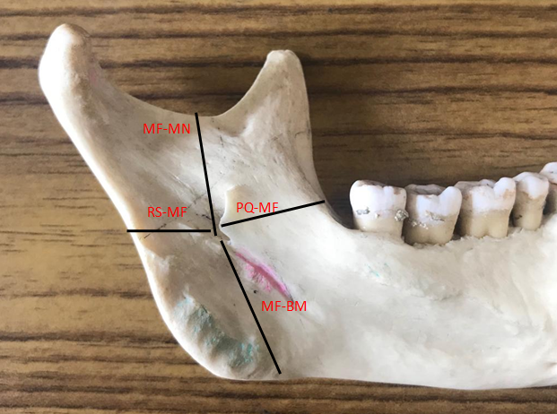
Distance from the midpoint of anterior margin of mandibular foramen to the nearest point on the anterior border of the ramus of mandible is denoted by PQ-MF.
Distance from the midpoint of posterior margin of mandibular foramen to the nearest point on the posterior border of the ramus of mandible is denoted by RS-MF.
Distance from the lowest point of mandibular notch to the inferior limit of mandibular foramen is denoted by MF-MN.
Distance from inferior limit of m andibular for amen to the base of the mandible is denoted by MF-BM.

Medial surface of mandibular ramus showing measurements taken from different landmarks to determine the position of the mandibular foramen.
PQ-MF: distance from the midpoint of anterior margin of mandibular foramen to the nearest point on the anterior border of the ramus of mandible
BM -M F: dsistance from inferior limit of mandibular foramen to the base of the mandible
MF-MN: distance from the lowest point of mandibula r notch to the inferior limit of mandibular foramen;
RS-MF: distance from the midpoint of posterior margin of mandibular foramen to the nearest point on the posterior border of the ramus of mandible.

All the above parameters were carefully measured and statistically analysed in the department of anatomy. Student’s t test was used as test of significance to compare the mean values of right and left sides. P-value less than 0.05 is statistically significant. The results of the present study were compared with the results of previous studies.
Results
A total of 100 adult human mandibles were studied for the location of mandibular foramen. The minimum, maximum, average and standard deviation values of the different parameters were studied on both the sides of the mandible and are shown in [Table 1]. There was no significant difference statatically between the values obtained on the right and left sides (P>0.05).
| Measurement | Right side (mm) | Left side (mm) |
| PQ-MF | 17.05 ± 2.70 | 17.35±3.00 |
| RS-MF | 10.41±2.13 | 9.72±2.06 |
| PQ-RS | 31.66±3.77 | 31.33±3.88 |
| BM-MF | 22.20±3.28 | 25.30±4.2 |
| MF-MN | 21.50±2.81 | 21.88±3.31 |
PQ-MF: distance from midpoint of anterior margin of mandibular foramen to the nearest point on anterior border of ramus.
RS-MF: distance from midpoint of posterior margin of mandibular foramen to the nearest point on posterior border of ramus.
BM-MF: mandibular notch to inferior limit of mandibular foramen.
MF-MN: distance from Mandibular Base to inferior limit of mandibular foramen.
MF-MN: The sum of the distances PQ-MF and RS-MF.
Location of mandibular foramen in relation anteroposterior and superoinferior axis of the ramus of mandible:
The percentile of the distance from anterior border of ramus to midpoint of mandibular foramen in relation to the distance from anterior to posterior border of ramus on the right side is 56.73±3.44% and on the left side it is 62.2±2.32%. The percentile of distance MF-MN in relation to MF - MN+ MF-MB is 49.68±3.46% on the right side and 46.51±5.1% on the left side. There is no statistically significant difference in the location of mandibular foramen on the right and left sides according to value of P>0.05 in both the anteroposterior axis and superoinferior axis.
| Side | Anteroposterior localization% | Superoinferior localization % |
| Right | 56.66±3.47 | 49.61±3.49 |
| Left | 62.22±2.31 | 46.51±5.2 |
Discussion
The knowledge of position of mandibular foramen is of a great importance for many procedures in dentistry. Mandibular foramen is a key landmark which lies on the inner aspect of the ramus of mandible. It is intimately related with inferior alveolar nerve which is branch of posterior division of mandibular nerve passing through same foramen. Many clinical conditions mentioned above in the study requires nerve block. So its exact location on medial aspect in relation to various landmarks need to carry out study. In present study mandibular foramen is studied in relation to distance from anterior, posterior border of ramus, distance from mandibular notch and mandibular base. Variations in anatomical postion of mandibular foramen may result in failure of inferior nerve block anesthesia. Several studies have been done to locate its position with the same aims.
According to K. Thangavedu et al the mandibular foramen is located 19mm + 2.34 from coronoid notch of ramus, 2.75mm posterior to the midpoint of anteroposterior width of ramus, 5mm inferior to midpoint of condyle to the inferior border distance.
According to Shalini et al mean distance of mandibular foramen from anterior border of ramus is 17.11 + 2.74mm on right and 17.41 + 3.05mm on left, from posterior is 10.47 + 2.74mm on right and 9.68 + 2.03mm on left side. In her study author has also found accessory mandibular foramen in 32.36% of mandibles. According to her study knowledge of mandibular foramen is useful to many clinicians like dental surgeons, oromaxillofacial surgeons.
Seema Gupta et al studied to find out accessory lingual foramen to avoid injury to inferior alveolar nerve while operating dental surgeries and procedures.
Shaikh Amjad has mentioned the mean of MF-AB distance 15.6mm on right and 15.3mm on left sides. Mean of MF-PB distance is 12.0mm on right and 11.60mm on left side. In his study he has taken MF-MB distance which was 23.4mm and 22.9mm on right and left side respectively.
Rajkumari K et al has studied the morphology of MF with the same aim to avoid complication of mandibular nerve block. The result obtained by her study is very close to that of the present study.
Kilarkaji et al[1] in his study on middle east Asian mandibles found that the distance from mandibular foramen to anterior border of ramus was 18.5±1.9mm on right side and 18.5±2.0mm on left side.
Varsha Shenoy et al[2] in her study on mandibles of south Indian origin found that mandibularforamen was located at a distance of 16.1mm on right side and 16.3mm on left side from anterior border of ramus of mandible.
The present study distance from mandibular foramen to anterior border of ramus was 17.05mm on right side and 17.35 mm on left side, no signifcant difference between right and left side and thus result is similar to result of study by Oguz et al[3] Varsha Shenoy et al but differs from Kilarkaji et al[1] and Prado et al.
In the present study distance between mandibular foramen to posterior border was 10.41mm on right and 9.72mm on left, no significant difference between sides. Distance from mandibular notch to mandibular foramen was 21.50mm on right side and 21.88mm on left side without any significant side difference.
In the present study distance from mandibular foramen to base of mandible or inferior border was 22.20mm on right side and 25.30mm on left side which is comp arable with Dr. Amita Sarkar who studied 50 dry adult human mandibles and the distance of mandibular foramen to base of mandible was 24.80±3.00 on right side and 24.60±3.10 on left side.
Conclusion
The data may also be useful in reconstructive surgery and anthropological assessments. The present study gives knowledge of the position of mandibular foramen and provides useful information regarding site of inferior alveolar nerve block which is routinely used by detal surgeons in performing various procedures like tooth extraction, removal of ramus of mandible by maxillofacial surgeon.
Source of funding
None.
Conflict of interest
None.
References
- N Kilarkaji, S R Nayak, P Narayan, L V Prabhu. The location of the mandibular foramen maintains absolute bilateral symmetry in mandibles of different age groups. Hong Kong Dent J 2005. [Google Scholar]
- Varsha Shenoy, S Vijayalakshmi, P Saraswathi. Osteometric analysis of the mandibular foramen in dry human mandibles. J Clin Diagnostic Res 2012. [Google Scholar]
- O Oguz, M G Bzkir. Evaluation of the location of the mandibular and the mental foramina in dry, young, adult human male dentulous mandibles. . [Google Scholar]
